Biomarker for Cognitive Function in Alcoholism
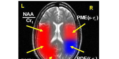
Brain magnetic resonance spectroscopic imaging (MRSI) studies examined 12 individual brain regions in cognitively intact (N = 4 males) and cognitively impaired (N = 5 males) chronic alcoholism subjects. Neuromolecular differences (p ≤ 0.0001) between the two groups are shown in the figure. Color intensity is scaled to the z-score (mean difference/standard deviation) which is a measure of the effect size of the differences. An increase in a molecular concentration is shown in red; a decrease is shown in blue.
The molecular changes associated with cognitive impairment in chronic alcoholism are increases in membrane phospholipid building blocks [PME(s-tc)] in the right superior temporal cortex and left inferior parietal cortex; increases in intermediates in phospholipid synthesis and/or lipid and protein glycosylation [(α-g)ATP] in the left occipital; a measure of neuronal numbers or functional integrity (NAA/Crt ratio) in the left superior temporal cortex and decreases in synaptic/transport vesicles [PDE(i-tc)] in the right inferior parietal cortex (Goldstein, Pettegrew, Cornelius, 2005).
